
A 6 mm Follicle on Day 2 of Your Period? Don’t Overlook This Detail Before IVF Egg Retrieval
A 6 mm Follicle on Cycle Day 2? Before IVF Egg Retrieval, Don’t Overlook This Detail
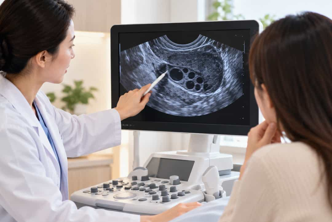
Many women preparing for pregnancy have had this experience: just on the second day of menstruation, when they go to the hospital for a baseline follicle check, ultrasound shows that one follicle has already reached 6 mm and is noticeably larger than the others.
Some people think this is a good thing, believing that if follicles grow quickly, ovarian function must be good. But for women preparing for IVF treatment, doctors often focus not only on how fast follicles grow, but also on whether the development of different follicles is coordinated.
If a clearly leading follicle appears early in the menstrual cycle, later ovarian stimulation may involve issues such as a widening size gap between follicles and inconsistent maturation times.
So why do follicles grow early? Does this mean egg quality is poor? For women preparing for third-generation IVF in Thailand, how can they tell whether the stimulation protocol needs adjustment?
Today, Huanqiu Xinsheng looks at this easily overlooked detail from three perspectives: follicle development patterns, hormonal changes, and assisted reproduction management.
1. Why has a follicle already reached 6 mm just as menstruation begins?
Follicle development does not suddenly start on the first day of menstruation; it is a process that spans different menstrual cycles and is continuously regulated by hormones.
Normally, multiple follicles at different developmental stages exist in the ovaries. After entering the early menstrual phase, as follicle-stimulating hormone (FSH) changes, a cohort of antral follicles continues to grow, and some gradually gain a developmental advantage.
During a baseline ultrasound, doctors usually observe the number, size, and distribution of antral follicles in both ovaries to provide reference information for later fertility assessment.
Note that antral follicles are not required to all be exactly the same size. A 6 mm follicle found on cycle day 2 is not necessarily abnormal.
What truly deserves attention is whether this follicle is clearly larger than the others, whether it is accompanied by abnormally elevated estradiol, and whether subsequent monitoring shows persistent asynchrony in development.
In clinical practice, the appearance of a clearly leading follicle early in the menstrual cycle is sometimes called “early follicular development.” However, this is not an independent disease name with unified diagnostic criteria.
For women preparing for IVF, simply seeing the number 6 mm does not mean you should immediately conclude that there is a problem with egg quality. Doctors need to consider follicle number, hormone levels, and previous stimulation response to further analyze its clinical significance.
2. Why do follicles develop early? The key is hormonal regulation
To understand early follicle growth, it helps to first understand hormonal changes during the menstrual cycle.
After ovulation, the original follicle turns into the corpus luteum and secretes progesterone and estradiol. These hormones not only participate in the secretory transformation of the endometrium but also affect hormone release from the hypothalamus and pituitary through feedback regulation.
As the corpus luteum gradually regresses, estradiol and progesterone levels decline, FSH begins to change, and conditions are created for the next cohort of follicles to continue developing.
If there is a timing difference in hormonal changes at this stage, some follicles may gain a growth advantage earlier, causing differences in follicle size early in the menstrual cycle.
However, early follicle growth cannot simply be attributed to luteal insufficiency.
Some women may have diminished ovarian reserve, elevated baseline FSH, or altered follicle recruitment rhythms. Especially with increasing age, the ovaries’ response to hormonal stimulation may change, and some follicles may be more likely to enter the dominant growth phase early.
In addition, normal fluctuations occur between different menstrual cycles. An occasional finding of a relatively large baseline follicle does not indicate that the endocrine system has a disease.
Therefore, doctors usually need to make a comprehensive judgment based on menstrual regularity, AMH, baseline FSH, LH, estradiol, and ultrasound results, rather than concluding that luteal function is abnormal based on a single hormone being high or low.
3. If follicles grow quickly, does it really mean poor egg quality?
This is the question many women preparing for pregnancy care about most.
First, it is important to clarify that there is no simple corresponding relationship between follicle size and egg quality.
A follicle is the microenvironment in which the oocyte grows and develops, containing follicular fluid, granulosa cells, and other supporting structures. As the follicle develops, the oocyte must complete complex nuclear maturation and cytoplasmic maturation processes before it can have the potential for further fertilization and embryo development.
Follicle diameter can help doctors determine the developmental stage, but it cannot directly reflect the chromosomal status or actual developmental capacity of the oocyte.
For women trying to conceive naturally, even if follicle growth is relatively fast in a given menstrual cycle, it does not mean that ovulation or conception cannot occur normally in that cycle.
For IVF patients, the situation requires further analysis.
If one follicle is clearly ahead while other follicles are still at a smaller stage, asynchronous follicle development may occur during subsequent stimulation.
When the leading follicle has already reached a size suitable for triggering final maturation, other follicles may not yet be fully developed; if treatment waits for the smaller follicles to continue growing, changes in the leading follicle also need to be considered.
This increases the complexity of determining the timing of the trigger injection and may also affect the final number of mature oocytes obtained.
But it should be emphasized that asynchronous follicle development does not necessarily cause egg retrieval failure, and there is insufficient evidence to prove that a 6 mm follicle on cycle day 2 directly increases the risk of oocyte chromosomal abnormalities.
Factors that truly affect treatment outcomes also include a woman’s age, ovarian reserve, stimulation protocol, oocyte maturation, and laboratory culture conditions.
4. Why does IVF stimulation pay particular attention to follicle synchrony?
A natural menstrual cycle usually centers on forming a dominant follicle and completing ovulation, whereas the goal of IVF ovarian stimulation is to obtain an appropriate number of mature oocytes while ensuring safety.
This requires coordinating the development pace of multiple follicles as much as possible.
During stimulation, doctors dynamically adjust gonadotropin dosage based on ultrasound monitoring results and hormonal changes, and select an appropriate time to trigger final oocyte maturation.
If there are obvious differences in follicle size, some smaller follicles may not yield mature oocytes at retrieval, thereby affecting the number of mature eggs.
However, clinical practice does not require all follicles to be exactly the same. A certain degree of size variation is very common, and doctors will make judgments based on the development of the overall follicle cohort.
For women with diminished ovarian reserve, a low number of eggs retrieved previously, or previous marked uneven follicle development, follicle synchrony is particularly worth attention.
Some patients may need adjustments to the start time of stimulation, drug dosage, or cycle management to improve follicle recruitment and growth.
It should be noted that improving synchrony does not mean all follicles can be made to mature, nor does it guarantee obtaining more high-quality embryos.
In addition, follicle development and endometrial changes are both affected by hormones, but a relatively large baseline follicle does not mean that the endometrium and embryo are directly out of sync.
Especially in treatment plans using frozen embryo transfer, the timing of embryo transfer can be arranged independently according to subsequent endometrial preparation.
Therefore, asynchronous follicle development and embryo implantation failure cannot simply be equated.
5. If early follicle development recurs, what tests are needed?
If a clearly leading follicle is found early in menstruation for several consecutive cycles, or if previous IVF treatment repeatedly showed large differences in follicle size, the following assessment areas can be discussed with a reproductive doctor.
1. Ovarian reserve and baseline hormones
AMH, baseline antral follicle count (AFC), and early menstrual FSH, LH, estradiol, and other indicators can help doctors understand ovarian reserve and the hormonal environment.
Among these, AMH is mainly used to predict ovarian response to stimulation drugs and cannot be used alone to judge egg quality.
If baseline estradiol is elevated, ultrasound is also needed to determine whether there is a functional cyst or other condition affecting hormone levels.
2. Menstrual cycle and post-ovulation hormonal changes
For women with markedly shortened menstrual cycles, a short luteal phase, or abnormal bleeding, doctors may further assess ovulation and luteal phase changes.
Progesterone testing can help determine whether ovulation has occurred, but progesterone secretion is markedly pulsatile, and a single blood test result cannot accurately evaluate the function of the entire luteal phase.
At present, diagnostic criteria for luteal insufficiency and some treatment measures remain controversial. It is not recommended to use progesterone on your own based solely on a single progesterone result.
3. Previous stimulation response and proportion of mature eggs
For women who have already undergone IVF treatment, previous cycle records are often of important reference value.
Doctors usually pay attention to the number of stimulation days, drug dosage, follicle growth curve, trigger timing, number of eggs retrieved, and proportion of mature oocytes.
If there were many follicles but few mature eggs obtained, or if there was a large developmental gap between follicles, this information can help doctors assess whether subsequent cycles need adjustment.
4. Age and overall health status
A woman’s age, ovarian reserve, weight changes, thyroid disease, and other endocrine problems may all affect menstruation and ovulation.
Long-term insufficient sleep and psychological stress may also affect reproductive endocrine regulation, but stress cannot be considered a direct cause of early follicle development on this basis.
The purpose of standardized assessment is to identify medical problems that truly need to be addressed, rather than continuously adding tests without clear necessity.
6. If follicles are asynchronous, can the IVF stimulation protocol be adjusted?
For patients with obvious asynchronous follicle development, reproductive doctors may consider different cycle management approaches based on the specific situation.
For example, some patients may receive pretreatment such as oral contraceptives or GnRH agonists before stimulation to regulate follicle recruitment and the hormonal environment.
Other patients may be suitable for a GnRH antagonist protocol directly, with medication adjusted during stimulation according to follicle growth.
For patients with diminished ovarian reserve or previous poor response, doctors also need to consider whether pretreatment may cause excessive suppression and whether it will increase treatment time.
Therefore, there is no uniform protocol suitable for all patients.
Especially for women over 35, with declining AMH, or with previous unsatisfactory egg retrieval results, the importance of age and treatment timing should not be ignored solely in pursuit of consistent follicle size.
It should also be noted that oral contraceptives, stimulation drugs, and hormonal drugs should all be used by doctors according to medical indications. It is not recommended to adjust the menstrual cycle or take medication in advance on your own.
7. Preparing for third-generation IVF in Thailand: how can cycle management be done in advance?
For families planning to travel to Thailand for assisted reproductive treatment through overseas fertility and cross-border reproductive services, understanding their own follicle development in advance can help improve the efficiency of cross-border medical arrangements.
If previous examinations found relatively large follicles early in the menstrual cycle, uneven follicle development, or a low number of mature eggs, it is advisable to organize relevant materials before traveling to Thailand.
These include recent AMH, baseline hormones, antral follicle count, menstrual cycle records, and previous IVF stimulation protocols and egg retrieval results.
Based on this information, doctors can determine whether further examination is needed, whether stimulation arrangements should be adjusted, or whether necessary management should be carried out before formally entering the treatment cycle.
It should be specifically noted that third-generation IVF in Thailand involves preimplantation genetic testing (PGT), which is mainly used for genetic evaluation in patients who meet medical indications. It cannot solve the problem of asynchronous follicle development, nor can it directly improve egg quality.
Therefore, ovarian assessment and stimulation management before treatment remain important parts of the assisted reproduction process.
Huanqiu Xinsheng focuses on assisted reproduction-related services in Thailand. Centered on the practical needs of families seeking fertility treatment overseas, it provides overseas medical accompaniment support such as hospital appointment coordination, organization of medical records, interpretation and accompaniment, transfer arrangements, accommodation guidance, and coordination of treatment steps.
For clients with previous asynchronous follicle development, diminished ovarian reserve, or repeated stimulation experiences, we can assist in organizing examination reports and treatment records, provide interpretation and communication during consultations, and help clients accurately understand doctors’ recommendations and follow-up arrangements.
All medical decisions involving stimulation protocols, hormonal medication, egg retrieval timing, and embryo transfer should be made by reproductive medicine professionals with appropriate qualifications.
Through full-cycle fertility management services, Huanqiu Xinsheng hopes to make cross-border medical communication smoother and ensure that every treatment stage is connected in an orderly way.
Conclusion: Fast follicle growth is not the problem; the key is the overall developmental rhythm
Finding a 6 mm follicle on cycle day 2 does not mean egg quality is necessarily poor, nor does it mean IVF treatment is destined to fail.
Follicle development itself varies between individuals. What truly deserves attention is the overall growth of the follicle cohort, the hormonal environment, and whether obvious asynchrony repeatedly occurs in previous cycles.
For women trying to conceive naturally, an occasional relatively large follicle usually does not require excessive intervention; for women preparing for assisted reproductive treatment, doctors should determine whether treatment strategy needs adjustment based on ovarian reserve and stimulation records.
Scientific fertility management is not about pursuing every follicle growing at the same speed, but about choosing a treatment plan that suits you on the basis of fully understanding your physical condition.
Huanqiu Xinsheng adheres to the concept of full-cycle fertility management and provides professional interpretation, accompaniment, and cross-border medical coordination support for families planning to receive IVF treatment in Thailand, making the path to overseas fertility treatment clearer and more orderly.
Huanqiu Xinsheng | Full-Cycle Fertility Management
Warm reminder: This article is for assisted reproductive health education only and does not constitute individualized medical advice. Follicle size cannot be used alone to judge egg quality. Specific examinations, medication, and stimulation protocols should be developed by professional reproductive doctors according to actual conditions.

- Previous
- Next